
Cervical Spine

The cervical spine is the neck region of the spinal column. It is composed of 7 bones (C1-C7) and runs from the base of your skull to the bottom of your neck. The cervical spine is very important, as it helps support the head, as well as allows for flexibility in bending the neck and turning the head from side to side.
Cervical Spinal Column
The cervical spinal column is made up of 7 bones, C1-C7. The 7 vertebrae are stacked on top of each other in order from the base of the skull (C1) to the bottom of the neck (C7). However two of the cervical vertebrae are different from the rest:
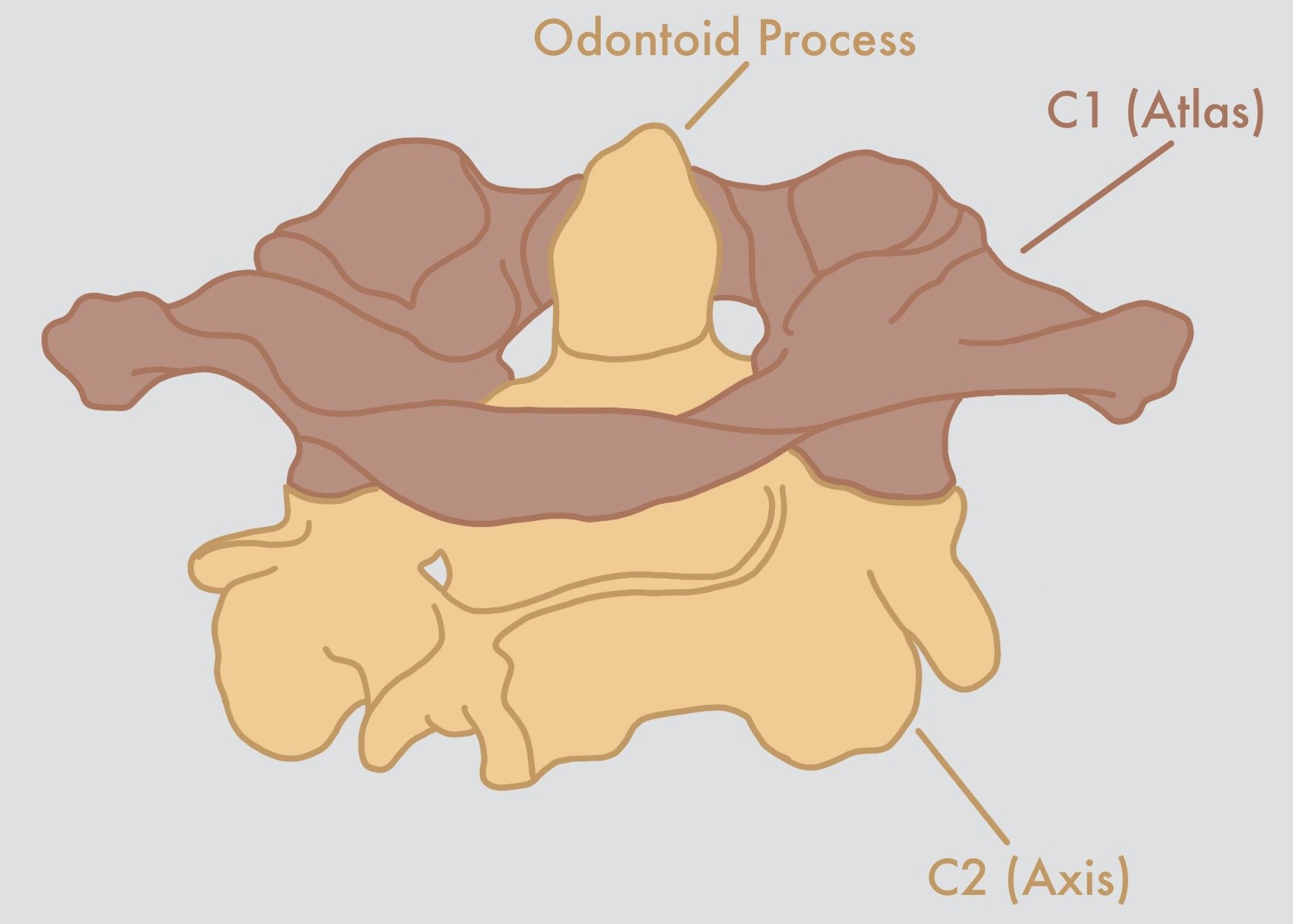
C1 (atlas): This vertebrae, located at the base of the skull, is a ring-shaped bone. Unlike other vertebrae in the spine, the atlas does not have a spinous process or a vertebral body.
Cervical Spinal Cord
The cervical spinal cord runs from the brainstem to the bottom of the C7 vertebrae. The main function of the cervical spinal cord is to communicate with the upper body (arms and hands).
C2 (axis): This vertebrae is responsible for the ability to turn our head from side to side. The axis has a round, bony projection called the odontoid process (also known as the “dens”), which sticks up into the circular hole of the atlas.


After C2, the rest of the cervical vertebrae have normal anatomy.
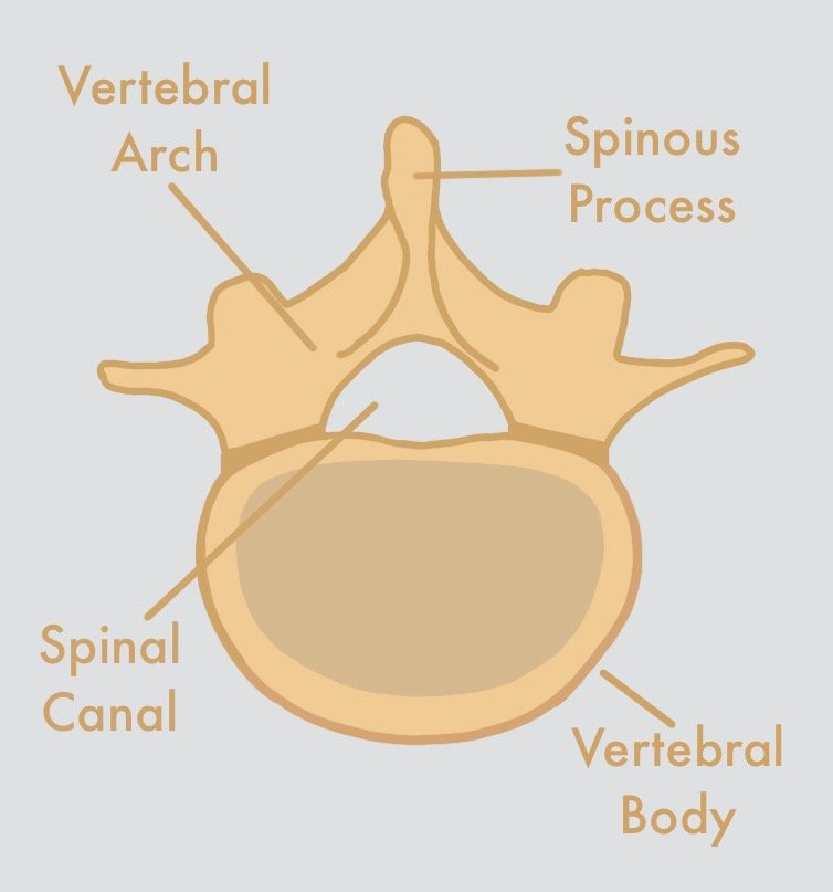
Each vertebrae is composed of a vertebral body and vertebral arch, which come together to create a hollow space within the vertebral column (spinal canal). On the back of each of the vertebrae is the spinous process, which are the bumps that you feel when you run your hand down the back of your neck.

Each cervical nerve communicates with a different part of the upper body, and is responsible for movement and sensation in that area. These areas, also called dermatomes, create a neurological map, which can be used by healthcare providers to detect and diagnose neurological disease.
X-ray of cervical spine
MRI of cervical spine